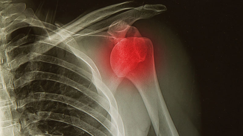
A shoulder dislocation occurs when the upper arm bone pops out of the shoulder socket, leading to extreme discomfort and impaired mobility. One of the key diagnostic tools for confirming a shoulder dislocation is an X-ray, which provides detailed images of the bones and joints involved.
In this article, we’ll explore the importance of a shoulder dislocation X-ray, how it aids in diagnosis and treatment, and what the various X-ray views can reveal. Whether you’re wondering what a dislocated shoulder looks like or seeking information about treatment planning, this comprehensive guide will answer all your questions.
What is a shoulder dislocation?

A shoulder dislocation happens when the ball-shaped head of the upper arm bone (humerus) slips out of its socket in the shoulder blade (scapula). This can occur due to traumatic injury, such as a fall or direct blow, and results in the shoulder losing its range of motion.
Different Types of Shoulder Dislocations
There are several types of shoulder dislocations, which depend on the direction the humerus moves:
- Anterior dislocation: The most common type, where the humeral head moves forward out of the socket.
- Posterior dislocation: Less common, occurring when the humeral head moves backward.
- Inferior dislocation: Rare, where the humeral head is displaced downward.
Each type of dislocation can result in different symptoms and complications, making proper diagnosis essential for appropriate treatment.
Signs and Symptoms of a Dislocated Shoulder
A dislocated shoulder can cause a variety of symptoms, ranging from pain to visible deformity. Some of the most common signs include:
- Severe pain: Especially when trying to move the shoulder.
- Deformity: The shoulder may appear out of place or visibly “off” compared to the other side.
- Limited mobility: A person with a dislocated shoulder may not be able to move their arm fully.
- Swelling and bruising: Inflammation is common following dislocation.
In rare cases, people may ask, “Can you dislocate your shoulder and not know?” While it’s uncommon, individuals with high pain tolerance or nerve damage might not feel the full effects immediately.
Why is an X-ray essential for shoulder dislocation diagnosis and treatment?
X-rays are a critical tool in diagnosing and treating shoulder dislocations. They not only confirm whether a dislocation has occurred but also provide vital information about the severity and type of dislocation.
Confirming the diagnosis
X-rays clearly show the position of the humeral head and whether it has moved out of the shoulder socket.
Assessing damage
X-rays can reveal additional fractures or bone injuries that may require special attention.
Planning treatment
The type and severity of the dislocation visible on the X-ray help physicians plan the most appropriate treatment approach.
How X-rays Work in Diagnosing Shoulder Dislocations

X-rays work by passing a controlled amount of radiation through the body, which produces images of the bones. For shoulder dislocations, X-rays are used to provide a clear view of the bones and joints in the shoulder area.
Capturing images from different angles
An X-ray of a dislocated shoulder is taken from different angles to ensure a comprehensive view of the dislocation. This is crucial because a single view might not capture the entire extent of the injury. Multiple angles, such as the anteroposterior (AP) view, scapular Y view, and axillary view, are typically used to provide a full picture of the joint and surrounding areas.
Identifying bone displacement
The X-ray images make it easy to see how far the humeral head has moved from its normal position. For example, in an anterior dislocation (the most common type), the humeral head will appear to be moved forward, out of the socket. Posterior and inferior dislocations show different patterns of displacement, which the X-ray will clearly reveal.
Checking for fractures
Shoulder dislocations can sometimes be accompanied by bone fractures, especially in older patients or those who experience severe trauma. The X-ray can reveal small cracks or full fractures in the humeral head or the shoulder socket itself. Detecting these fractures is essential, as they may require different treatment strategies compared to a simple dislocation.
One Step Diagnostic is a leading radiology center specializing in high-quality imaging services to provide accurate diagnoses for a variety of medical conditions, including shoulder injuries.
Benefits of Using X-rays for Shoulder Injuries
X-rays are invaluable when dealing with shoulder injuries because they offer several benefits. Their ability to provide clear, real-time images of the shoulder joint allows for accurate diagnosis and treatment planning.
Immediate diagnosis
One of the main benefits of X-rays is the speed at which they provide results. X-rays offer real-time imaging, which allows doctors to quickly determine whether a shoulder is dislocated and assess the severity of the injury. This rapid feedback is crucial for reducing the dislocation and preventing further damage to the shoulder’s soft tissues and nerves.
Non-invasive
X-rays are a non-invasive and painless diagnostic tool, making them ideal for patients with shoulder injuries. Unlike some imaging techniques that may require injections or more complex procedures, an X-ray involves simply positioning the patient and capturing the necessary images. This quick and easy process helps patients avoid unnecessary discomfort during an already painful injury.
Detailed imagery
X-rays provide highly detailed images of the bones and joints in the shoulder. They can reveal the exact position of the humeral head in relation to the shoulder socket, helping doctors identify not only the dislocation but also any associated fractures or bone damage. This level of detail is critical for determining the type of dislocation—whether anterior, posterior, or inferior—and for evaluating the best treatment options based on the severity of the injury.
Types of Shoulder Dislocation X-Ray Views
When diagnosing shoulder dislocations, different x-ray views are used to provide a comprehensive image of the injury. These include:
Anteroposterior (AP) view
The AP view shows the shoulder from front to back. This view provides a general overview of the shoulder joint and is often the first X-ray taken.
Scapular Y view
The scapular Y view offers a side image of the shoulder, making it easier to detect dislocations and fractures. It also highlights the alignment of the shoulder blade.
Axillary view
The axillary view captures the shoulder joint from below, providing critical information about the position of the humeral head relative to the socket.
What can the X-ray reveal about shoulder dislocation?

An X-ray shoulder dislocation image reveals several important aspects of the injury. Understanding “what does a dislocated shoulder look like” through X-ray imaging helps doctors create effective treatment plans.
- Type of dislocation: Whether it’s an anterior, posterior, or inferior dislocation.
- Associated fractures: Bone breaks or cracks may be visible.
- Severity: The degree of displacement of the humerus from the socket.
Treatment Planning Based on X-ray Results
Once an X-ray confirms the dislocation, treatment plans can be developed based on the specific type and severity of the injury. For minor dislocations, manual reduction may be enough, where the doctor repositions the bone into the socket. More severe cases might require surgery or immobilization followed by physical therapy.
Ensuring Accurate Diagnosis with Shoulder X-rays
Shoulder dislocations can be painful and significantly impact your range of motion. Using X-rays for diagnosis is an essential step in accurately identifying the type and severity of the injury. X-rays provide clear, detailed images of the bones and joints, which helps medical professionals develop a personalized treatment plan based on the dislocation’s specifics.
For those seeking expert diagnostic services, One Step Diagnostic, a trusted radiology center, offers comprehensive X-ray imaging to ensure accurate diagnosis and effective treatment planning. Whether you’ve experienced a shoulder injury or are in need of routine imaging, One Step Diagnostic provides the care and expertise necessary to get you back on the path to recovery. Contact them today!