Chronic pain can be challenging to diagnose and treat, as its causes are often hidden beneath the surface. This is where diagnostic imaging becomes essential. By providing a clear look inside the body, techniques like X-rays, MRIs, and CT scans help pinpoint the source of pain, from nerve damage to joint issues.
With these insights, doctors can create personalized treatment plans that target the exact cause, offering more effective and lasting relief for patients.
In this article, we’ll explore the critical role that diagnostic imaging plays in chronic pain management and how it can lead to better patient outcomes.
Importance of Accurate Diagnosis for Effective Treatment

Accurate diagnosis is key to addressing chronic pain effectively, as it helps healthcare providers tailor treatments specific to the underlying issue. With proper diagnostic tools, patients can avoid unnecessary treatments and begin the path to lasting relief.
- Targeted Treatment Plans: When the root cause of pain is identified, doctors can create personalized treatment strategies. This leads to faster recovery and better pain management, reducing trial-and-error methods. Treatment plans often involve a multidisciplinary approach, including physical therapy and, for some, services like online occupational therapy to help regain function and adapt daily routines to manage their condition effectively.
- Prevention of Further Damage: Diagnosing the cause of chronic pain early on helps prevent complications or worsening of the condition. Accurate diagnosis can prevent the need for more invasive treatments in the future.
- Improved Patient Confidence: Knowing the exact cause of pain reassures patients and gives them a clear direction for their treatment. This understanding improves outcomes, and many find that complementing their medical care with practices that support mental well-being, such as mindfulness or wearing crystal bracelets as a personal token, helps manage the stress of their health journey.
Understanding Chronic Pain
Chronic pain is pain that lasts for weeks, months, or even years, often outlasting the normal healing process. It can affect many aspects of a person’s life, limiting mobility, causing emotional distress, and decreasing quality of life. Common causes of chronic pain include:
- Arthritis: Inflammation in the joints that causes pain, stiffness, and reduced range of motion.
- Back Issues: Conditions like herniated discs, sciatica, and spinal stenosis can cause persistent back pain.
- Nerve Damage: Conditions like neuropathy, where nerves are damaged, leading to ongoing pain, often described as burning or stabbing.
- Fibromyalgia: A condition characterized by widespread musculoskeletal pain, fatigue, and tenderness in localized areas.
Types of Diagnostic Imaging for Chronic Pain

There are several diagnostic imaging for chronic pain that help healthcare professionals understand the source of chronic pain. These methods provide a detailed look at the body’s internal structures and are essential for accurate diagnosis and treatment planning.
MRI (Magnetic Resonance Imaging)
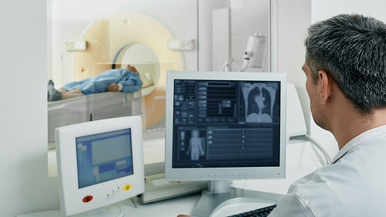
MRI for chronic pain diagnosis uses strong magnetic fields and radio waves to create detailed images of soft tissues, including muscles, ligaments, and nerves. This is particularly useful for detecting conditions like disc herniations, nerve compression, and other soft tissue injuries that may be contributing to chronic pain.
CT Scans (Computed Tomography)
CT scans for chronic pain combine X-ray images taken from different angles to create cross-sectional images of the body. This imaging technique is ideal for identifying bone fractures, joint abnormalities, and certain types of tumors or infections that may be causing pain.
X-rays
X-ray for pain management are commonly used to visualize bones and detect fractures, joint problems, or degenerative conditions such as osteoarthritis. While they are limited in showing soft tissue damage, they remain a primary tool in diagnosing skeletal issues that could be contributing to chronic pain.
Ultrasound
Ultrasound uses sound waves to produce images of soft tissues and organs in the body. It’s particularly useful for assessing soft tissue injuries like muscle strains, tendonitis, or inflammation in joints and is often used for real-time guidance in procedures like imaging-guided injections or fluid aspiration.
DEXA Scan (Dual-Energy X-ray Absorptiometry)
DEXA scans are primarily used to measure bone density and assess bone health, helping to identify osteoporosis or other bone-related issues. By detecting bone loss, this imaging method can help address pain caused by weakened bones or fractures, leading to more targeted treatment options.
Benefits of Diagnostic Imaging for Chronic Pain Patients

Diagnostic imaging provides numerous benefits to chronic pain patients, offering a clearer understanding of the underlying causes of pain and enabling more effective treatment strategies. These advanced tools not only enhance the accuracy of diagnoses but also help doctors tailor treatments that can lead to better long-term outcomes.
Accurate Diagnosis of Pain Sources
One of the primary benefits of diagnostic imaging is its ability to precisely identify the source of pain. By revealing details about the bones, joints, muscles, and nerves, imaging techniques help doctors pinpoint the exact cause of discomfort, ensuring that treatment targets the right issue and improves patient outcomes.
Personalized and Targeted Treatment Plans
With the insights gained from diagnostic imaging, doctors can create customized treatment plans based on each patient’s unique condition. This means that therapies, medications, and interventions are chosen specifically to address the identified cause of the pain, leading to more efficient and effective results.
Enhanced Effectiveness of Pain Management Techniques
By identifying the specific areas causing pain, diagnostic imaging helps doctors select the most appropriate pain management techniques, such as physical therapy, injections, or medication. This targeted approach can significantly increase the effectiveness of these treatments, providing patients with more significant relief from their chronic pain.
Reduction in the Need for Exploratory Surgery
Before the advent of diagnostic imaging, many patients underwent exploratory surgery to diagnose the source of pain. Today, imaging techniques like MRIs and CT scans allow doctors to view internal structures non-invasively, significantly reducing the need for risky and costly surgeries.
Continuous Monitoring and Adjustment of Treatments
Diagnostic imaging provides a way to continuously monitor the progression of a patient’s condition, ensuring that treatments remain effective. Regular imaging can show how well a patient is responding to therapy, allowing doctors to make timely adjustments to the treatment plan as necessary.
Early Detection of Complications or Secondary Conditions
Imaging techniques can detect complications or secondary conditions that may arise due to chronic pain, such as muscle atrophy or nerve damage. Identifying these issues early allows for quicker intervention, preventing further deterioration of the patient’s health and improving long-term outcomes.
Patient Reassurance and Improved Understanding of Pain
Diagnostic imaging offers patients a clearer understanding of their condition by providing a visual representation of the underlying causes of their pain. This not only reassures patients that their symptoms are valid but also empowers them to be more actively involved in their treatment journey.
What to Expect During an Imaging Appointment
Imaging appointments are typically straightforward and non-invasive, offering a detailed look at the source of your pain. While the process may vary depending on the type of imaging, you can generally expect a comfortable and efficient experience.
- Preparation: For some imaging tests, such as MRIs, you may be asked to avoid eating or drinking beforehand. You’ll also be instructed to remove any metal objects, as they can interfere with the imaging process.
- Duration: Most imaging procedures take between 20 to 60 minutes, depending on the type of test and the area being examined. For example, an X-ray takes only a few minutes, while an MRI may take longer.
- Positioning: You will be asked to lie down or sit in a specific position during the scan. The technician may need to adjust you for the best angle to capture clear images of the affected area.
- Results: After the procedure, a radiologist will analyze the images and provide a report to your doctor. They will then discuss the findings with you and recommend any further treatment or next steps.
Choose One Step Diagnostic for Top-Notch Service
At One Step Diagnostic, we are committed to providing the highest quality diagnostic imaging services to help you get to the bottom of your chronic pain. Our experienced technicians and advanced technology ensure accurate results from our chronic pain diagnostic tests that lead to effective treatment. Take the first step towards pain relief—schedule your appointment with One Step Diagnostic today!


