Blood clots can be silent threats. They can lurk within the body, and without any warning signs, they can obstruct blood flow, leading to severe health complications, or even life-threatening situations if they remain undetected and untreated. It’s this silent nature that makes understanding and employing the right diagnostic tools so crucial for early detection and timely intervention.
Among various diagnostic methods, Magnetic Resonance Imaging (MRI) is a popular choice. But people sometimes ask: does an MRI show blood clots? Let’s dive into this together. In this article, we’re going to explore what MRI is, how it works, and whether it’s up to the task of finding blood clots.
Importance of Detecting Blood Clots

Detecting blood clots is crucial for several reasons, all of which revolve around preventing serious health issues and saving lives. Here’s why it’s so important:
- Preventing Blockages
Blood clots can block blood flow to vital organs. For instance, a clot in the arteries of the heart (coronary artery) can lead to a heart attack, while a clot that travels to the brain can cause a stroke. Early detection allows for timely treatment to dissolve or remove the clot, restoring normal blood flow and preventing organ damage.
- Avoiding Complications
Certain types of blood clots, such as deep vein thrombosis (DVT), can lead to long-term complications like post-thrombotic syndrome, characterized by pain, swelling, and in severe cases, ulcers in the affected limb. Early treatment can reduce the risk of these complications.
- Reducing the Risk of Pulmonary Embolism
DVT clots can break loose and travel to the lungs, causing a pulmonary embolism (PE), a potentially fatal condition. Detecting clots early in the deep veins can prevent PE by managing the clot before it becomes a greater threat.
- Saving Lives
Simply put, early detection and treatment of blood clots save lives. Whether it’s preventing a stroke, heart attack, or pulmonary embolism, the timely identification of blood clots allows for interventions that can avert fatal outcomes.
- Guiding Treatment Plans
Detecting a blood clot helps healthcare providers determine the best course of treatment, which may include anticoagulants (blood thinners), thrombolytics (clot dissolvers), or in some cases, surgical intervention. The nature and location of the clot play a crucial role in deciding the treatment strategy.
Understanding MRI: What It Is and How It Works
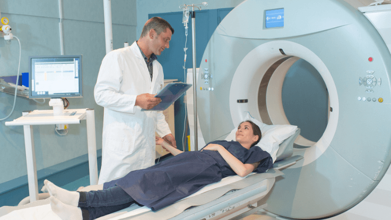
Magnetic Resonance Imaging (MRI) is a non-invasive diagnostic technique that produces detailed images of the internal structures of the body. Using a combination of a powerful magnetic field, radio waves, and computer technology, MRI scans can visualize organs, soft tissues, bone, and virtually all other internal body structures without the use of ionizing radiation (X-rays). Here’s how a step-by-step process of how it works:
- Definition and Preparation
An MRI scan is designed to capture high-resolution images of the inside of the body. Before the scan, patients are asked to remove any metal objects to avoid interference with the magnetic field. In some cases, a contrast agent may be administered to enhance the clarity of the images.
- Magnetic Field Activation
The patient lies inside the MRI machine, which then generates a strong magnetic field around them. This magnetic field temporarily realigns hydrogen atoms in the body, which are abundant due to the water content in tissues.
- Radiofrequency Pulses
Once the magnetic field is stable, the machine emits short bursts of radio waves toward the area being examined. These waves knock the aligned hydrogen atoms out of position.
- Energy Emission
As the hydrogen atoms return to their original alignment, they emit energy. This energy release varies between different types of tissues based on their chemical composition and physical properties.
- Signal Collection
The MRI scanner detects the energy emitted by the hydrogen atoms and relays this information to a computer. The nature and intensity of these signals differ based on the type of tissue and its condition, allowing the machine to distinguish between various tissue types.
- Image Processing
The computer processes the signals to create detailed images. Each signal corresponds to a specific slice of the body, enabling the creation of cross-sectional images from different angles. These slices can be compiled to generate comprehensive 3D images of the scanned area.
- Analysis
Finally, a radiologist reviews the images to identify any abnormalities, such as tumors, inflammation, or injury. The precision of MRI allows for the detailed examination of organs and soft tissues, facilitating accurate diagnoses and informing treatment decisions.
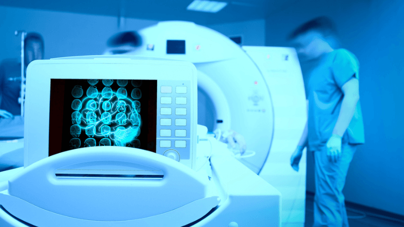
Can MRI Detect Blood Clots?

Yes, MRI can detect blood clots. MRI is particularly useful for visualizing soft tissues and the vascular system, areas where blood clots can pose significant risks. This includes spotting clots tucked away in tricky spots like the brain, spinal cord, or deep within veins
The process involves using MRI’s powerful magnetic field and radio waves to create images that can show the presence of abnormal blood flow or blockages indicative of clots. Specific MRI techniques, such as Magnetic Resonance Venography (MRV) or Magnetic Resonance Angiography (MRA), are designed to image the body’s veins and arteries, offering detailed views that can help in identifying clots.
It is worth noting that while MRI is highly effective in detecting blood clots in certain areas, it’s not the first-line diagnostic tool for all types of clots. For example, ultrasounds are often used for detecting deep vein thrombosis (DVT) in the legs due to their accessibility and cost-effectiveness. However, for complex cases or when clots are suspected in less accessible areas of the body, MRI provides a non-invasive, detailed examination method without the exposure to ionizing radiation.
How MRIs Detect Blood Clots
MRI detects blood clots through a sophisticated process that leverages its ability to create detailed images of the body’s internal structures. Here’s a closer look at how this technology zeroes in on blood clots:
- Creating Contrast in Tissues
MRI is exceptional at differentiating between the various types of tissues within the body. This is crucial because blood clots, depending on their stage and location, can appear differently from the surrounding tissues. MRI can highlight these differences, making clots more visible.
- Visualizing Blood Flow
Using techniques like MRV (Magnetic Resonance Venography) for veins and MRA (Magnetic Resonance Angiography) for arteries, MRI can visualize blood flow—or the lack thereof—within the body. Clots create areas where blood flow is obstructed, and these disruptions can be seen on MRI images as blockages or abnormalities in the normal flow patterns.
- Detecting Abnormalities in Vascular Structures
Blood clots often cause changes in the shape or size of blood vessels. MRI can detect these structural abnormalities, indicating the presence of a clot. This is particularly useful for identifying clots in deep veins or in parts of the body where other imaging methods might not be as effective.
- Leveraging Contrast Agents
In some cases, a contrast agent might be used to enhance the visibility of blood vessels and clots on MRI scans. These agents improve the contrast between the clot and surrounding tissues, making it easier to identify and assess the clot.
- Utilizing Advanced Imaging Sequences
MRIs can use different imaging sequences to focus on specific types of tissue or abnormalities. Some sequences are particularly effective at highlighting blood clots, based on the properties of the clot and the surrounding tissues.
Through these steps, MRI provides a comprehensive view that not only shows the presence of a clot but can also give insights into its size, location, and potentially its impact on surrounding tissues.
Strengths of MRI in Blood Clot Detection
MRI represents a cornerstone in medical imaging, particularly for its precision in detecting blood clots. Let’s delve into the core strengths that elevate MRI’s role in identifying blood clots:
Superior Image Resolution
MRI’s hallmark is its capacity to generate exceptionally detailed images. This precision is critical for accurately identifying blood clots, even those that are small or located in complex anatomical regions.
Unmatched Soft Tissue Contrast
One of MRI’s most notable advantages is its excellent ability to differentiate between various types of soft tissues. This capability ensures that blood clots can be distinctly visualized against the backdrop of surrounding tissues.
Absence of Ionizing Radiation
Unlike CT scans and X-rays, MRI does not rely on ionizing radiation. This makes it a safer alternative for patients, particularly when multiple scans are necessary over time. This reduces any long-term health risks associated with radiation exposure.
Versatile Imaging Techniques
MRI can utilize a range of imaging techniques, such as Magnetic Resonance Venography (MRV) and Magnetic Resonance Angiography (MRA). This is tailored to visualize the vascular system in detail. These specialized techniques are adept at highlighting blood clots within veins and arteries, offering a clear view of potential blockages.
Functional and Anatomical Insights
Beyond merely showing the physical presence of a clot, MRI can also provide valuable information about the effect of a clot on blood flow and the surrounding tissues. This functional insight aids in assessing the severity of the clot and planning the most effective treatment strategy.
MRI Limitations and Considerations
While MRI is a powerful tool for detecting blood clots and offering detailed insights into the body’s internal structures, it does come with its own set of limitations and considerations. Understanding these is key to maximizing its benefits while navigating its drawbacks effectively. Here are some important points to consider:
Accessibility and Cost
MRI scanners are sophisticated pieces of equipment that are not as widely available as other diagnostic tools, like ultrasound machines. Additionally, the cost of an MRI scan can be significantly higher, which might not be feasible for all patients or healthcare settings.
Time-Consuming Scans
An MRI scan can take a considerable amount of time, often ranging from 30 minutes to an hour or more. This can be challenging for patients who have difficulty remaining still for extended periods, including young children or those with claustrophobia.
Metal Interference
The strong magnetic field of the MRI machine means that patients with certain metal implants, such as pacemakers, cochlear implants, or some metal clips and screws from previous surgeries, may not be eligible for an MRI scan due to safety concerns or the risk of distorting the images.
Contrast Agent Reactions
In some cases, a contrast agent is used to enhance the clarity of MRI images. While rare, there can be allergic reactions or side effects to these agents, requiring careful screening and monitoring by medical professionals.
Claustrophobia and Discomfort
The enclosed nature of traditional MRI machines can cause feelings of claustrophobia or discomfort in some patients. Although open MRI options exist, they may not be available in all facilities and can sometimes result in lower-quality images.
Not Always the First Choice
For certain types of clots, such as those in the legs (deep vein thrombosis), ultrasound might be preferred due to its immediacy and effectiveness. MRI is often reserved for more complex cases or when other imaging modalities fail to provide adequate information.
Interpretation Requires Expertise
The detailed images produced by MRI require interpretation by experienced radiologists. The complexity of MRI images means that accurate reading and diagnosis depend on high levels of skill and experience.
MRI vs Other Diagnosis Methods
When comparing MRI to other diagnostic methods for detecting blood clots and various medical conditions, it’s essential to weigh their strengths, limitations, and best use cases. Each method brings its own set of advantages to the table, making the choice highly dependent on the specific clinical scenario.
MRI vs. Ultrasound
- Ultrasound is often the first line of investigation for diagnosing blood clots, especially in the legs (deep vein thrombosis, DVT) and in pregnancy. It’s quick, portable, and cost-effective, offering real-time images of blood flow and clot formation without radiation. However, its effectiveness can be limited by the operator’s skill and the patient’s body composition.
- MRI, on the other hand, provides superior detail, especially for soft tissue contrast and in areas that are difficult to visualize with ultrasound, like the pelvis, abdomen, and brain. While more expensive and time-consuming, MRI is invaluable when detailed imaging is necessary to guide treatment decisions.
MRI vs. CT Scan
- CT Scans are highly effective for quickly visualizing blood clots in the lungs (pulmonary embolism) and for assessing bone injuries. They offer faster scanning times compared to MRI and are widely available in emergency settings. However, CT scans use ionizing radiation, which can be a concern with repeated exposure.
- MRI excels in cases where soft tissue detail is crucial, such as in neurological conditions, joint disorders, and certain types of vascular clots. It’s preferred when a more detailed examination is required without the risks associated with radiation exposure.
MRI vs. X-Ray
- X-Rays are best suited for evaluating bone fractures and lung and heart conditions but offer limited information on soft tissues and are not typically used for detecting blood clots directly.
- MRI provides a comprehensive view of both hard and soft tissues, making it a better choice for conditions where soft tissue detail is necessary for diagnosis.
MRI vs. Venography
- Venography involves injecting a contrast dye directly into the veins to visualize blood clots, particularly in the legs. It’s invasive and rarely used today due to the availability of less invasive, equally effective options.
- MRI, with techniques like Magnetic Resonance Venography (MRV), offers a non-invasive alternative to traditional venography, providing detailed images of the veins without the need for contrast injection into the veins, though intravenous contrast may still be used in some MRI examinations.
Navigating Towards Health with One Step Diagnostic
MRI can truly play a crucial role in detecting blood clots, offering a safe and detailed imaging option. However, it is part of a broader diagnostic toolkit, with its use tailored to specific cases. For those seeking expert diagnostic services, including MRI, One Step Diagnostic stands ready to provide comprehensive care with cutting-edge technology.
Discover more about how One Step Diagnostic can guide you towards a healthier future by visiting our services page or contacting us here. Remember that early detection and the right tools, like MRI, are key to managing health risks effectively. Trust in specialists like One Step Diagnostic to take the right steps towards safeguarding your health.