For athletes and active individuals, injuries are often part of the journey. Physical setbacks can be frustrating, from a twisted knee during a game to lingering muscle pain after a workout. Some injuries heal with rest and rehab, while others require a deeper look to understand what is going on beneath the surface.
This is where Magnetic Resonance Imaging, or MRI, proves to be invaluable. When it comes to diagnosing soft tissue damage, joint instability, or hidden stress fractures, sports injuries and MRIs go hand in hand. This guide explores how MRIs work, which injuries they detect, and why they are often the first choice for medical professionals in sports medicine.
What is an MRI and How Does it Work?
MRI, short for Magnetic Resonance Imaging, is a powerful diagnostic tool that uses magnetic fields and radio waves to create detailed images of the body. It is particularly useful for examining muscles, ligaments, tendons, cartilage, and joints.
The machine detects signals from hydrogen atoms within the body and translates them into high-resolution images. This level of detail allows physicians to evaluate the severity and nature of an injury accurately. In the context of MRI for sports injuries, it is one of the most reliable ways to examine soft tissues that cannot be seen clearly with other imaging techniques.
Common Sports Injuries Diagnosed by MRI
Athletes often experience a range of injuries. An MRI can help pinpoint the exact location and extent of the damage to guide effective treatment.
Muscle & Tendon Injuries
- Muscle strains & tears: Muscle injuries are frequent among athletes. Minor strains may heal quickly, but full tears require more intensive care. An MRI provides the best imaging for muscle tears, revealing the depth of the injury and helping plan a proper recovery.
- Tendonitis & tendon ruptures: Tendonitis develops through repetitive stress, while ruptures may happen suddenly. An MRI can identify inflammation or torn fibers, giving doctors the information needed to decide on conservative treatment or surgery.
Ligament Injuries
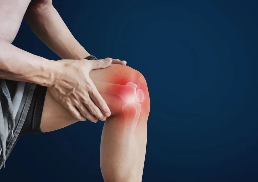
- ACL, MCL, PCL tears: The knee is especially vulnerable in sports. A torn ligament MRI scan helps confirm the presence and severity of tears in ligaments like the ACL or MCL, which is crucial for treatment planning.
- Ankle sprains & ligament damage: Sprains may seem minor, but repeated injuries can lead to chronic instability. MRI scans are often used to assess the condition of ankle ligaments after swelling or instability persists.
Bone & Joint Injuries
- Stress fractures: These injuries are subtle and often missed by X-rays. MRI detects them in early stages, which helps prevent further damage.
- Cartilage tears: Cartilage in joints can wear down or tear from impact or overuse. MRI helps detect these issues early, allowing for quicker intervention.
- Joint dislocations & instability: After a joint dislocation, an MRI is used to examine surrounding ligaments and cartilage to assess any additional injury.
Overuse & Chronic Conditions
- Shin splints (MTSS): Medial tibial stress syndrome causes pain along the shinbone. MRI is used to rule out more serious conditions and confirm the diagnosis.
- Plantar fasciitis: MRI can show thickening or degeneration of the plantar fascia, which often causes persistent heel pain.
- Bursitis & tendon degeneration: Inflamed bursae or worn tendons contribute to chronic discomfort. MRI reveals swelling and tissue breakdown that may not appear on other scans.

Why MRI is the Best Choice for Sports Injuries
When comparing MRI vs X-ray for sports injuries, MRI provides a far more detailed view of soft tissues and hidden damage. It is considered the preferred method for comprehensive diagnosis.
Superior soft tissue detail
MRI provides detailed images of muscles, ligaments, tendons, and other soft tissues. This level of clarity helps medical professionals diagnose injuries that standard X-rays often miss.
No radiation exposure
MRI uses magnetic fields and radio waves to generate images instead of ionizing radiation. This makes it a safer option for athletes, especially when multiple scans are needed throughout treatment.
Helps guide surgical planning
Detailed MRI scans give surgeons a clear view of the injured area, including soft tissue and joint structures. This information is essential for planning procedures that require precision and accuracy.
Detects hidden injuries
An MRI can reveal internal injuries like microtears, fluid buildup, or early stress fractures that are not visible on other imaging tests. This ensures athletes receive appropriate care before the condition worsens.
What to Expect During a Sports Injury MRI
Understanding what the sports injury diagnosis MRI process involves can help ease any anxiety athletes may have before a scan.
Preparation
Most MRIs do not require special preparation. Patients are usually asked to remove all metal objects and wear comfortable clothing. Any medical implants should be disclosed to the technician.
Procedure
The scan typically lasts between 30 to 60 minutes. The patient will lie still while the machine takes multiple images. Staying still is important to ensure image clarity.
Contrast dye use
In some cases, a contrast dye is injected to highlight specific areas or abnormalities. This helps improve the accuracy of the scan, especially when evaluating joints or blood flow.
Post-scan process
Once the scan is complete, a radiologist will review the images and send a report to the referring physician. Results usually come within a few days. Most patients can return to normal activity right after the appointment.

When should athletes get an MRI?
Timing is important in diagnosing and treating injuries. Here are some situations when an MRI is highly recommended.
Persistent pain after initial treatment
If pain lingers despite physical therapy or medication, an MRI can reveal underlying issues that may not have been detected initially. This helps doctors make more informed decisions and adjust the treatment plan accordingly.
Suspected ligament/cartilage tears
Injuries with sudden pain, swelling, or joint instability often indicate torn ligaments or damaged cartilage. An MRI provides a clear view of these internal structures to confirm the extent of the damage.
Pre-surgical evaluation
Before surgery, an MRI helps map out the injured area in detail, giving surgeons valuable insight. This ensures a more precise and successful surgical approach.
Unexplained swelling or limited mobility
When swelling or stiffness continues without an obvious cause, MRI can uncover hidden injuries such as fluid buildup or small tears. Early detection through imaging leads to quicker and more effective treatment.
Your Road to Recovery Starts with the Right Imaging
This guide has shown how sports injuries and MRIs are closely linked. MRI offers superior imaging for soft tissue, bone, and joint conditions. It is safe, precise, and extremely valuable for both diagnosis and treatment planning. From muscle strains to ligament tears and chronic overuse conditions, MRI remains one of the most effective tools in modern sports medicine. Staying informed about when to use an MRI can make all the difference in your recovery journey.
For athletes seeking top-tier imaging services, One Step Diagnostic is a trusted provider in Texas. With advanced MRI technology, fast scheduling, and expert radiologists, we ensure a smooth and professional experience from start to finish. Schedule your MRI today!


