Soft tissue injuries, such as muscle tears, ligament sprains, and tendonitis, are common issues that many individuals face. Whether from sports, accidents, or overuse, these injuries can significantly affect daily life. To accurately assess and treat these conditions, healthcare providers use imaging tools, with ultrasound for soft tissue injuries standing out as a top choice.
This article explores why doctors recommend ultrasound when dealing with soft tissue injuries. You’ll learn about the imaging process, the types of injuries it detects, the benefits of ultrasound for muscle tear diagnosis, and how it plays a key role in treatment planning. We’ll also guide you through what to expect during the procedure and where to go for high-quality diagnostic services.
What Are Soft Tissue Injuries?
Soft tissue injuries refer to damage involving muscles, tendons, ligaments, or fascia. These injuries can be caused by acute trauma, repetitive strain, or improper movement. Common types include strains, sprains, contusions, tendonitis, and bursitis. Symptoms usually involve swelling, pain, restricted mobility, and bruising.
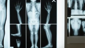
Because soft tissues don’t appear clearly on X-rays, soft tissue injury diagnosis ultrasound has become a preferred method for evaluating the extent of damage. Ultrasound offers high-resolution images that allow healthcare providers to pinpoint the specific injury and make informed treatment recommendations.
How Ultrasound Works
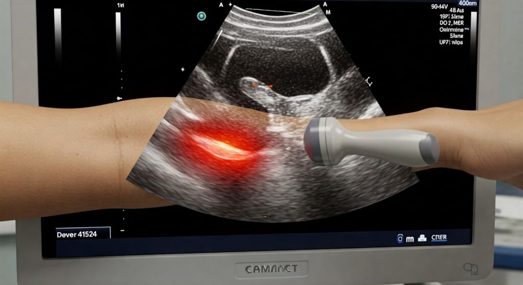
Ultrasound for soft tissue injuries uses high-frequency sound waves to create live images of muscles, ligaments, and tendons beneath the skin. A technician uses a small handheld device called a transducer, which sends sound waves into the body. The waves bounce back from tissues and are translated into real-time visuals on a monitor.
Unlike CT scans or MRIs, ultrasound is free of radiation, making it ideal for repeated use and safe for a wide range of patients. For soft tissue injury diagnosis, ultrasound enables clinicians to visualize injuries as they happen, especially when the body part is moved during scanning. This dynamic imaging gives a more comprehensive understanding of functional impairments.
Key Advantages of Ultrasound for Soft Tissue Injuries
Here are some key advantages of ultrasound for soft tissue injuries:

Real-Time Imaging
One of the most significant advantages of ultrasound for soft tissue injury diagnosis is its ability to provide real-time imaging. This allows doctors to observe how soft tissues, such as muscles or ligaments, are functioning during movement. This dynamic capability helps in assessing the severity of injuries like muscle tears and tendon strains more effectively than static imaging methods.
Excellent Soft Tissue Detail
Ultrasound for soft tissue injuries provides clear, detailed images of the affected area. Unlike X-rays, which primarily show bones, ultrasound captures the texture, structure, and integrity of muscles, tendons, and ligaments. This detailed view aids doctors in identifying specific damage or abnormalities that may not be visible through other imaging methods.
Non-Invasive and Safe
Another reason why doctors recommend ultrasound is that it’s a non-invasive procedure. There is no need for incisions or injections, and the procedure is entirely safe. Since ultrasound uses sound waves instead of radiation, it is suitable for people of all ages, including children and pregnant women.
Relatively Inexpensive and Accessible
Compared to other imaging techniques, such as MRI or CT scans, ultrasound for soft tissue injuries is generally more affordable and widely available. Its cost-effectiveness makes it an attractive option for diagnosing soft tissue injuries without the need for expensive equipment or lengthy procedures.
Portable
Ultrasound machines are portable and can be moved from one location to another. This mobility means that ultrasound can be used in various settings, including clinics, sports facilities, and emergency rooms, providing convenience and accessibility when immediate diagnosis is required.
What Ultrasound Can Reveal About Soft Tissue Injuries
Ultrasound is incredibly versatile and can reveal a wide range of injuries and conditions related to soft tissues. Here are some common types of soft tissue injuries that ultrasound can detect:
Muscle Tears and Strains
Ultrasound for muscle tear diagnosis is essential for determining the extent of damage to muscle fibers. It can identify both partial and complete tears, helping doctors create a treatment plan to promote healing and prevent further injury.
Tendonitis and Tendinosis
Tendonitis (inflammation of a tendon) and tendinosis (degeneration of a tendon) are common injuries that ultrasound can easily detect. It allows doctors to assess the severity of the inflammation and determine the best approach for treatment, such as physical therapy or injections.
Ligament Sprains and Tears
Ultrasound is highly effective in detecting ligament sprains and tears, particularly in areas like the ankle, knee, and shoulder. By revealing the extent of the tear or strain, ultrasound helps doctors assess the need for surgical intervention or conservative treatment.
Bursitis
Bursitis, which occurs when the fluid-filled sacs around joints become inflamed, is another condition that ultrasound can reveal. Ultrasound helps doctors visualize the inflamed bursa and evaluate the amount of fluid accumulation, guiding treatment options.
Hematomas and Fluid Collections
When soft tissues are injured, blood can accumulate and form hematomas, or fluid-filled cysts can develop. Ultrasound for soft tissue injuries can detect these fluid collections and assist in determining if drainage or other treatments are necessary.
Foreign Bodies
Sometimes, foreign objects such as splinters or debris can become embedded in soft tissues. Ultrasound is an excellent tool for locating these foreign bodies and helping doctors remove them safely.
Nerve Entrapment (in some cases)
In certain cases, ultrasound can reveal nerve entrapment, which occurs when a nerve is compressed by surrounding tissues. This condition can cause pain, numbness, and tingling sensations, and ultrasound helps identify the underlying issue.
How This Information Aids Diagnosis and Treatment
Ultrasound for soft tissue injuries provides valuable information that aids in both diagnosis and treatment. Here’s how:
- It allows doctors to determine the precise location and extent of the injury.
- It helps identify the specific tissue affected (e.g., muscle, tendon, ligament).
- It guides decisions about treatment methods, such as rest, physical therapy, or surgery.
- It provides insight into the healing process, allowing doctors to monitor progress and adjust treatments as needed.

What to Expect During an Ultrasound Examination for a Soft Tissue Injury
If you’re scheduled for an ultrasound for soft tissue injuries, here’s what you can expect during the examination:
- You’ll be asked to lie down in a comfortable position, and the affected area will be exposed.
- A gel will be applied to the skin to help the transducer make better contact with the body.
- The technician will move the transducer over the injury site, which may feel slightly cool or pressure but is generally painless.
- The images generated will be analyzed by a radiologist or doctor, who will then discuss the results with you.
The Role of Ultrasound in Soft Tissue Injury Diagnosis and Treatment
Ultrasound for soft tissue injuries plays a vital role in diagnosing and treating muscle tears, tendon injuries, ligament sprains, and more. Its real-time imaging, non-invasive nature, and affordability make it a valuable tool for doctors, ensuring accurate diagnosis and optimal treatment plans for patients.
If you’re in need of an ultrasound for a soft tissue injury, consider visiting One Step Diagnostic for fast, reliable imaging services. With experienced professionals and state-of-the-art equipment, One Step Diagnostic is dedicated to providing you with the best care possible to help you recover quickly and effectively. Contact us today to schedule your ultrasound appointment!